Compared with standard-resolution (SR) PCCTA, ultrahigh-resolution (UHR) PCCTA achieved clearly stronger correlation with invasive FFR and higher diagnostic accuracy for functionally significant stenosis.
UHR-PCCTA-derived FFR maintained performance in heavily calcified and diffusely diseased vessels, where SR-PCCTA-derived FFR did not show significant correlation with invasive FFR.
Agreement with invasive hemodynamic classification was better for UHR-PCCTA than for SR-PCCTA, supporting the value of ultrahigh-resolution imaging for coronary CTA-based FFR assessment.
Access publication
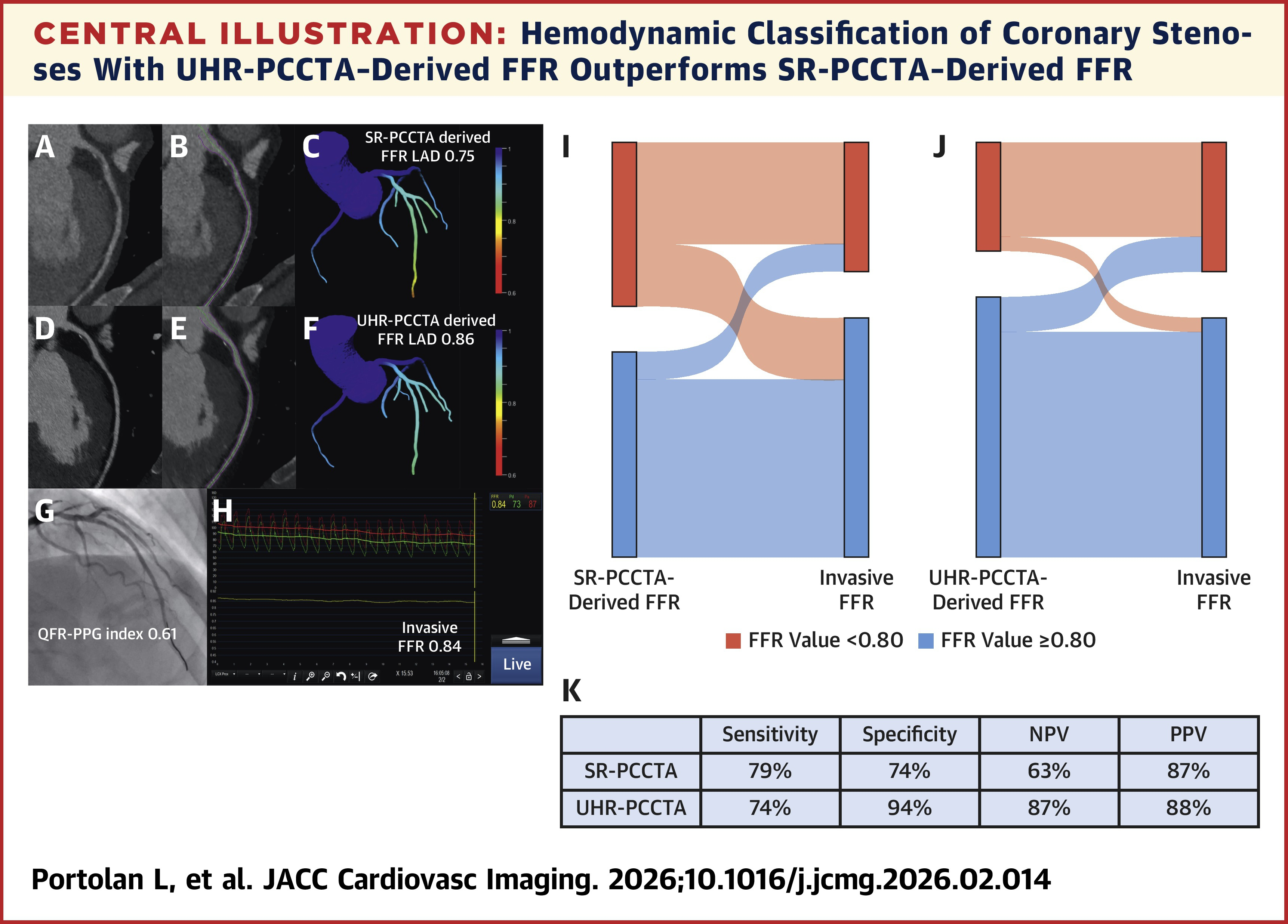
Central illustration: UHR-PCCTA derived FFR with invasive FFR as a reference (I, J) with higher specificity, negative predictive value, positive predictive value (K). Illustrative example of SR-PCCTA–derived FFR (A to C) and UHR-PCCTA–derived FFR (D to F) of an intermediate stenosis of the LAD using invasive FFR as a reference (G, H). FFR = fractional flow reserve; ICA = invasive coronary angiography; LAD = left anterior descending; NPV = negative predictive value; PCCTA = photon-counting coronary computed tomography angiography; PPV = positive predictive value; QFR-PPG = quantitative flow ratio–pullback pressure gradient; SR = standard resolution; UHR = ultrahigh resolution.
Source: Portolan et al., JACC: Cardiovascular Imaging, 2026.
Protocol
Scanner | NAEOTOM Alpha |
Software | VA50 SP1 |
Scan area | Coronary CTA / heart |
Scan mode | SR-PCCTA and UHR-PCCTA |
Scan direction | n.a. |
Tube voltage [kV] | 140 SR | 120 UHR |
Effective mAs [mAs] | n.a. |
IQ level | 89 SR | 69 UHR |
Dose modulation | CARE Dose4D |
CTDIvol [mGy] | n.a. |
DLP [mGy*cm] | 91 median SR | 296 median UHR |
Effective dose [mSv] | 2.6 median SR | 8.3 median UHR |
Rotation time [s] | 0.25 |
Pitch | n.a. |
Matrix | n.a. |
Slice collimation [mm] | 144 x 0.4 SR | 120 x 0.2 UHR |
Slice width [mm] | 0.4 SR | 0.2 UHR |
Reconstruction increment [mm] | n.a. |
Reconstruction kernel | Qr48 SR | Qr56 UHR |
Iterative reconstruction | QIR 4 |
keV level [keV] | 67 for SR-PCCTA-derived FFR analysis |
Contrast agent | Omnipaque 350 mgI/mL |
Contrast volume | 70 mL SR | 80 mL UHR |
Flow rate | 4.5 mL/s |
Saline chaser | 50 mL |
ECG technique | Mainly prospective triggering; retrospective gating in selected patients |
Acquisition phase | Mostly diastolic; systolic only in selected SR cases |
Heart rate control | Sublingual nitroglycerin 400-800 μg; IV metoprolol up to 40 mg targeting <60 bpm |
Portolan L, Kotronias RA, Andreaggi S, et al. Standard and Ultrahigh Resolution Photon-Counting Coronary CTA-Derived FFR Against Invasive FFR Assessment. JACC: Cardiovascular Imaging. Just Accepted. Published March 25, 2026.
